self pay ultrasound pricing
General Body
Abdominal Ultrasound Complete
This scan includes liver, gallbladder, pancreas, right and left kidneys, common bile duct, spleen, aorta
Abdominal Ultrasound Limited
This scan evaluates one or multiple organs.
Abdominal Aorta Ultrasound
This test used visually to show an aneurysm in the main artery leaving the heart which supply blood to the abdomen, pelvic and legs
Renal Ultrasound Complete
This scan includes both kidneys and bladder with post void residual. (Urine)
Bladder Ultrasound
This test evaluate the size of bladder when full and measure the bladder when empty to see how much post void residual (urine) was retain in the bladder along with any other abnormalities.
Kidneys Only
A kidney ultrasound is performed to measure the kidneys to make sure they aren't enlarged, to look for stones, masses, fluid collection, cyst, infection and other disorders.
Transabdominal Prostate Ultrasound
This test evaluate the bladder, prostate gland size and seminal vesicles. Most of the common symptoms "not being able to urinate: having a hard time stopping or starting the flow of urine; needing to urinate often, especially at night; weak flow of urine, urine flow that starts and stop; pain or burning upon urination.
Cardiac And Vascular
Echocardiogram
This is a sonogram of the heart which shows the size and shape of the heart, pumping capacity, location and extent of any tissue damage.
Carotid Artery Ultrasound
A scan that help doctors determine stroke risk by measuring the flow of blood through the carotid arteries that supply blood to the brain.
Renal Artery Ultrasound
These arteries supply oxygenated blood to your kidneys. This test help to detect blockages or narrowing of the arteries.
Upper Or Lower Extremities Arteries
An arterial exam of the legs or arms that looks at the blood circulation in the arteries to see if there is any blockage.
Upper or Lower Extremities Venous
This exam is to search for blood clots, especially in the veins in the legs and long standing leg swelling.
Upper or Lower Uni-lateral Extremity Vein
An exam that check for a clot in the vein of one extremity.
Upper or lower Uni-lateral Extremity Artery
An exam that check for a blockage in the artery of one extremity.
Small Parts Ultrasound
Ultrasound procedures are a useful way of visualizing trauma, swelling, cysts, nodules & other abnormalities of many superficial location of the body such as the thyroid gland, neck, breasts, groin and the testes.
Breast Ultrasound
Ultrasound scan is used to help diagnose breast lumps or other abnormalities during a physical exam or on a Mammogram or breast MRI.
Uni-Lateral (single breast $243)
Testicular Ultrasound
This exam determine enlargement of one or more of the testicles, painful testicles and blood flow in the testicles.
Thyroid Ultrasound
An Imaging method used in diagnosis of tumors, cysts or goiters and the size and number of nodules on your thyroid.
Female Pelvic Ultrasound
In women, a pelvic ultrasound is most often performed to evaluate the uterus, cervix, ovaries, fallopian tubes, bladder. Non- pregnant.
Transabdominal Pelvic Complete
This exam is done externally and create pictures of the organs inside your pelvis - the area between your belly and legs. This test can help diagnose problems like tumors or cysts.
Transvaginal ultrasound
Examines the reproductive organs from inside the vaginia. Scan can show up changes in your womb, ovaries or surrounding structures. Also common indication for checking placement of an IUD.
Pelvic Complete and Transvaginal
The combined use of transabdominal ultrasound and transvaginal ultrasound is clinically proven and therefore, medically necessary when either study is insufficient to provide adequate diagnosis.
1st Trimester ultrasound
4 weeks early ultrasound $152
6 weeks early ultrasound $152
6 weeks early ultrasound $152

In the early stages of pregnancy, the ultrasound scan will be performed via the vagina. (transvaginal ultrasound) This type of ultrasound provides a clearer view of the uterus and establish pregnancy confirmation
6 weeks early ultrasound $152
6 weeks early ultrasound $152
6 weeks early ultrasound $152

The Transvaginal ultrasound scan, however, should be able to confirm gestational age and due date. See the baby's heartbeat, location in uterus, yolk sac, the number of fetuses.
8 weeks early ultrasound $152
6 weeks early ultrasound $152
8 weeks early ultrasound $152

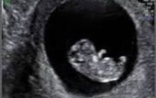
This ultrasound scan shows a little figure that looks something like an oblong bean. Eyes on the face and eyelids have formed. Limb buds are visible. Looks more like a human.
Second Trimester Ultrasound $252
Fetal Anatomy Scan
This exam use a transducer that is placed on your abdomen to view your baby, Anatomy scan about 18 to 22 weeks into you pregnancy, sex of your baby, how many fetus you are carrying and other findings.
Third Trimester Ultrasound $202
This is ultrasound after 28 weeks, commonly much later. It may also be referred to as a growth scan or late pregnancy ultrasound.
Fetal Anatomy Scan during the third trimester will be $352
Some examples of reasons to have a third trimester ultrasound are:
* Fetal position and size
* Placenta position and maturing
* Movement decrease
* AFI (amniotic Fluid Measurement)
* Weight
* The fetus or the mother's abdomen, feels too big or small for the stage of pregnancy
* A fetal abnormality was noted at an earlier ultrasound and requires follow-up
Add a footnote if this applies to your business
SKIN AND SOFT TISSUE INFECTION
Skin Abscess $250
Skin Abscess $250
Skin Abscess $250

Bacteria may get inside the elbow from an injury, insect bite or puncture wound break in the skin.
SOFT TISSUE MASS ULTRASOUND
Ganglion cyst $250
Popliteal (Baker's) cyst $250
Popliteal (Baker's) cyst $250

A non cancerous fluid filled lump on the tendons or joint of wrists and hands
Popliteal (Baker's) cyst $250
Popliteal (Baker's) cyst $250
Popliteal (Baker's) cyst $250

A fluid filled lump behind the knee that causes pain, bruising and limit mobility.
Lipoma $250
Popliteal (Baker's) cyst $250
Angiolipoma $250

Round or oval - shaped lump of fat tissue that grows just beneath the skin.
Angiolipoma $250
Epidermoid Cyst $250
Angiolipoma $250

A small soft tumor made up of fatty cells and blood vessels. Found in the head, neck and forearm.
Epidermoid Cyst $250
Epidermoid Cyst $250
Epidermoid Cyst $250
.jpg/:/cr=t:0%25,l:0%25,w:100%25,h:100%25/rs=w:388,h:194,cg:true)
Small lumps that develop on the head, neck, back or genitals
Musculoskeletal Ultrasound
Knee Effusion $250
Gout Bursitis of the foot $250
Gout Bursitis of the foot $250

Knee effusion is called water on the knee. Occurs when excess water accumulates in or around the knee joint. Common causes arthritis and injury cartilage in knee
Gout Bursitis of the foot $250
Gout Bursitis of the foot $250
Gout Bursitis of the foot $250

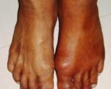
Affect the joints. Causing inflammation and severe pain
Glass Foreign body in hand $250
Wood foreign body in hand $250
Wood foreign body in hand $250

Pain at the location of the glass splinter. Redness, swelling, warmth or pus. A small speck or line under the skin.
Wood foreign body in hand $250
Wood foreign body in hand $250
Wood foreign body in hand $250

Feeling the presence of an object lodge in skin. Pain, bleeding, inflammation and infection.
FERTILITY ULTRASOUND $250

Ovary Follicular count
This type of scan is a series of ultrasound vaginal scans used to identify if a woman is ovulating and pinpoint when a follicle rupture and release an egg. Ovarian antral follicles can be identified and counted using transvaginal ultrasound. (A probe that is placed inside the vagina)
3D/4D Babyface Ultrasound
Gender Reveal $85
3D/4D Babyface $85
Gender Reveal $85

Prince or Princess
Can't wait to find out?
We will be glad to tell you if you are 15 weeks. 30 minute session
Peace of mind $85
3D/4D Babyface $85
Gender Reveal $85

Feeling Anxious, prior miscarriage.
Listen to heartbeat, observe baby's movements and check baby's position. 15 minute sessions
3D/4D Babyface $85
3D/4D Babyface $85
3D/4D Babyface $85

Can be done anytime of pregnancy. Optimal time 30-35 weeks
Gender upon request
30 minute session
Copyright © 2024 Happy Heart Imaging Service - All Rights Reserved.
Powered by GoDaddy Website Builder